The management of localized prostate cancer has evolved beyond a strict choice between active surveillance and radical, whole-gland treatment. For decades, men with intermediate-risk or localized disease had two primary curative options: surgical removal of the prostate (radical prostatectomy) or whole-pelvis radiation therapy. While effective at eradicating cancer, both treatments treat the entire gland indiscriminately, carrying recognized risks of urinary incontinence, erectile dysfunction, and bowel irritation due to their proximity to delicate pelvic nerves, sphincters, and the rectum.
This clinical gap led to the emergence of focal therapy, an approach designed to destroy index tumor lesions while sparing the healthy remainder of the prostate gland, the surrounding neurovascular bundles, and the urinary sphincter. Leading this non-invasive paradigm is High-Intensity Focused Ultrasound (HIFU)—an advanced technology that uses targeted sound waves to heat and destroy cancer cells without a single surgical incision.
In South Florida, Dr. Mourad Abouelleil is a board-certified urologist with the Palm Beach Health Network Physician Group, serving patients at Good Samaritan Medical Center in West Palm Beach and Palm Beach Gardens Medical Center. Dr. Abouelleil earned his medical degree from the University of Wisconsin School of Medicine and Public Health and completed his general surgery and urology residency training at SUNY Upstate Medical University in Syracuse, New York. Specializing in robotic surgery, minimally invasive urology, and targeted ablation, Dr. Abouelleil utilizes image-guided HIFU to deliver precise cancer control while helping men protect their urinary continence and sexual potency.
The Mechanism of Action: How HIFU Destroys Malignant Tissue
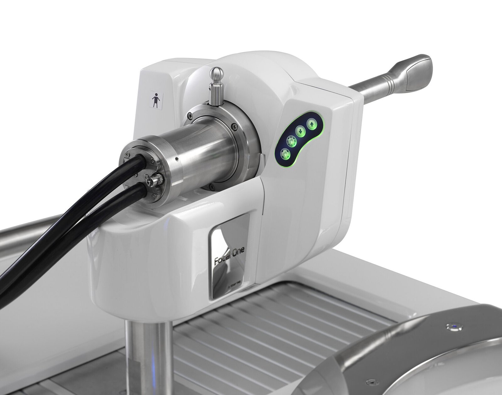
High-Intensity Focused Ultrasound operates on a principle similar to sunlight focused through a magnifying glass. Standard diagnostic ultrasound uses low-energy sound waves that bounce off organs harmlessly to create an image. HIFU, by contrast, uses a high-energy transducer that converges multiple intersecting acoustic beams into a single, tightly defined focal point.
- Coagulative Thermal Necrosis: At the focal point—typically measuring only a few millimeters in size—the rapid absorption of acoustic energy raises tissue temperatures to 85°C to 90°C (185°F to 194°F) within seconds. This intense heat denatures cellular proteins and causes instant coagulative thermal necrosis, destroying the targeted tumor tissue.
- Acoustic Cavitation: The mechanical vibration of high-frequency ultrasound waves creates microscopic vapor bubbles within intracellular water. As these micro-bubbles rapidly expand and collapse under acoustic pressure, they produce mechanical shear waves that disrupt cancer cell membranes.
- Precise Boundary Margin: Tissue lying outside the narrow focal zone—such as the rectal wall, the bladder neck, and the external urethral sphincter—remains completely unaffected because the individual sound waves pass through intervening structures at low, harmless energy densities.
The Role of Multiparametric MRI and Fusion Guidance
The success of focal HIFU depends on knowing the precise anatomical coordinate of the cancer lesion. Rather than treating blind, modern HIFU relies on Multiparametric MRI (mpMRI) Fusion Guidance:
- Preoperative Diagnostic MRI: A detailed 3-Tesla prostate MRI identifies the “index lesion” (the dominant tumor driving oncologic risk) and confirms whether it is confined within the prostatic capsule.
- Real-Time Transrectal Fusion: In the operating room, the high-resolution MRI data is digitally fused with real-time transrectal ultrasound (TRUS) imaging.
- Sub-Millimeter Contouring: The urologist outlines the exact treatment volume on a 3D computerized map. Safety margins are drawn around the tumor, while critical exclusion zones are marked around the neurovascular bundles (controlling erections) and the membranous urethra (controlling urination).
- Automated Robotic Delivery: The robotic HIFU arm delivers thousands of overlapping focal pulses throughout the contoured zone, systematically ablating the targeted lesion slice by slice.
Treatment Strategies: Focal vs. Hemi-Ablation vs. Whole-Gland
Depending on tumor volume, location, and the patient’s prostate anatomy, Dr. Abouelleil customizes the ablation boundary:
- Focal (Targeted) Ablation: Only the proven cancerous lesion and a small rim of healthy margin tissue are ablated. This approach offers the highest rates of potency and continence preservation and is ideal for isolated, unilateral index lesions.
- Hemi-Ablation: The entire lateral half (lobe) of the prostate harboring the tumor is ablated, while the contralateral half containing healthy tissue and its intact neurovascular bundle is spared.
- Whole-Gland HIFU: The entire prostate gland is treated. This is generally reserved for men who cannot undergo surgery or radiation due to age or medical comorbidities, or as a secondary salvage option following recurrent disease.
Comparative Overview: Radical Prostatectomy vs. Radiation vs. Focal HIFU
| Clinical Feature |
Radical Prostatectomy (Robotic) |
External Beam Radiation (EBRT) |
Targeted Focal HIFU |
| Surgical Invasiveness |
Abdominal keyhole incisions |
Non-invasive (External beams) |
Zero incisions (Transrectal probe) |
| Tissue Treated |
Entire prostate + seminal vesicles |
Entire prostate + margins |
Targeted lesion + safety boundary |
| Urinary Continence |
Temporary leakage; gradual recovery |
Lower initial incontinence; late toxicity |
Immediate or early control preserved |
| Erectile Function Preservation |
Dependent on nerve-sparing plane |
Gradual decline over 2–3 years |
High preservation (spares bundles) |
| Hospital Stay |
1 to 2 days inpatient |
Daily outpatient visits over weeks |
True outpatient (Go home same day) |
| Catheter Requirement |
5 to 7 days |
Rarely needed |
Temporary catheter for 3 to 7 days |
| Future Treatment Options |
Salvage radiation available |
Salvage surgery is highly complex |
Surgery or radiation remain fully open |
Patient Candidacy: Who is an Ideal Candidate for HIFU?
HIFU is an advanced therapeutic tool, but proper patient selection is essential to achieve oncologic success:
- Ideal Candidates:
- Men with localized, organ-confined prostate cancer (Stage T1c to T2a).
- Favorable intermediate-risk or select low-risk disease (Gleason 3+4=7 or low-volume 4+3=7).
- Concordant disease: The tumor visible on multiparametric MRI matches the location of positive biopsy cores.
- Patients seeking to avoid the sexual and urinary side effects associated with radical surgery or radiation.
- When HIFU May Not Be Recommended:
- Large anterior tumors located beyond the reach of the ultrasound focal depth ($> 40\text{ mm}$ from the rectal probe).
- Significant prostate calcifications that cause acoustic shadowing and deflect ultrasound energy.
- High-risk, aggressive, or metastatic disease (Gleason 8–10, PSA $> 20\text{ ng/mL}$, or bone/nodal spread) requiring systemic or broad multimodal therapy.
Recovery Trajectory and Post-Procedure Life
Because HIFU involves no abdominal incisions, muscle cutting, or blood loss, the recovery timeline is straightforward:
- Outpatient Discharge: The procedure is performed under general anesthesia and typically takes 1.5 to 2.5 hours. Patients recover in the post-anesthesia care unit and return home the same day.
- Catheter Care: Due to temporary swelling of the prostate following thermal ablation, a temporary Foley catheter or suprapubic tube is kept in place for 3 to 7 days to allow post-treatment edema to subside.
- Return to Activity: Most men resume light walking, driving, and desk work within 48 to 72 hours. Strenuous exercise and heavy lifting can typically be resumed after two weeks.
- Long-Term Monitoring: Because a portion of the prostate gland remains intact, patients follow a structured surveillance protocol involving PSA testing every three months, a follow-up multiparametric MRI at 6 to 12 months, and targeted biopsy if indicated.
Conclusion
A diagnosis of localized prostate cancer does not always require surgical removal of the entire gland or broad pelvic radiation. Through image-guided High-Intensity Focused Ultrasound (HIFU), Dr. Mourad Abouelleil provides men with a non-invasive, outpatient pathway that targets the cancer at its source while protecting the urinary control and sexual vitality that define quality of life.
- Surgeon: Dr. Mourad Abouelleil, MD
- Board Certification: American Board of Urology
- Practice Group: Urology Medical Associates – Palm Beach Health Network Physician Group
- Hospital Affiliations: Good Samaritan Medical Center (West Palm Beach) & Palm Beach Gardens Medical Center
- Primary Office Locations:
- West Palm Beach: 1411 North Flagler Drive, Suite 3800, West Palm Beach, FL 33401
- Jupiter: 641 University Boulevard, Suite 105, Jupiter, FL 33458
- Port St. Lucie: 900 Southeast Becker Road, Port St. Lucie, FL 34984
- Office Consultations: (561) 291-7182