
When a solid kidney mass is detected on an abdominal CT or MRI, the primary surgical objective is achieving complete cancer eradication. Historically, the default approach for renal tumors was radical nephrectomy—the complete removal of the affected kidney, surrounding fat, and adrenal gland. While oncologically sound, long-term clinical data revealed an unintended consequence: removing an entire kidney doubles the risk of developing chronic kidney disease (CKD), which significantly elevates long-term risks of cardiovascular events, stroke, and overall mortality.
Partial Nephrectomy, also known as Nephron-Sparing Surgery (NSS), has become the reference standard of care for clinical T1 renal masses (tumors up to 7 centimeters). The operation removes only the malignant tumor along with a thin rim of normal tissue, leaving the remaining healthy kidney parenchyma intact and fully functional. However, when tumors are complex—deeply embedded within the kidney (endophytic), adjacent to major vascular trunks, or abutting the urinary collecting system—the procedure demands exceptional technical precision.
In South Florida, Dr. Mourad Abouelleil is a board-certified urologist with the Palm Beach Health Network Physician Group, operating at Good Samaritan Medical Center in West Palm Beach and Palm Beach Gardens Medical Center. Having completed his medical training at the University of Wisconsin and comprehensive general surgery and urology residency at SUNY Upstate Medical University in Syracuse, New York, Dr. Abouelleil utilizes advanced robotic-assisted platforms to resect complex, anatomically challenging renal tumors. By combining multi-articulated robotic instrumentation, intraoperative ultrasound mapping, and rapid parenchymal reconstruction, Dr. Abouelleil preserves critical kidney filtration while delivering complete oncologic margins.
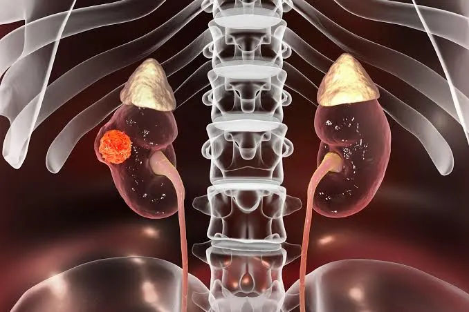
Each human kidney contains approximately one million microscopic filtering units called nephrons. Nephrons cannot regenerate once lost; their destruction is permanent. Preserving functional nephrons during cancer surgery yields profound systemic benefits:
Not all renal masses present the same surgical challenge. A superficial, outward-growing (exophytic) growth on the lower pole is relatively straightforward to remove, whereas a tumor buried deep within the renal parenchyma adjacent to the main renal artery and vein presents high operative complexity.
Dr. Abouelleil utilizes standardized scoring systems, such as the R.E.N.A.L. Nephrometry Score, to map tumor anatomy and guide the robotic approach:
Higher nephrometry scores (scores of 10 to 12) identify high-complexity tumors that previously were managed with complete kidney removal. With robotic multi-articulation and dynamic 3D optical magnification, many of these complex, deep-seated tumors can be resected while saving the kidney.
Robotic partial nephrectomy is a meticulously orchestrated procedure centered on a strict timeline to protect kidney tissue from oxygen deprivation:
Depending on whether the tumor is located on the anterior or posterior surface of the kidney, Dr. Abouelleil accesses the retroperitoneum or abdominal cavity through 4 to 5 small keyhole incisions (5 to 12 mm). Gerota’s fascia is incised, and the kidney is mobilized to clearly expose the target lesion.
The renal hilum is carefully dissected to isolate the main renal artery and vein. In select cases, Dr. Abouelleil isolates specific secondary (segmental) arterial branches directly feeding the tumor, allowing for selective regional clamping while leaving the remainder of the kidney continuously perfused.
A specialized, robotic ultrasound probe is introduced directly into the surgical field and controlled via the surgeon console. Sweeping the probe across the kidney surface allows the surgeon to visualize the hidden endophytic margins of the tumor beneath the renal cortex, locate feeding blood vessels, and verify the distance to the urinary collecting system.
To prevent heavy bleeding during resection, temporary micro-vascular bulldog clamps are placed on the renal artery, pausing blood flow (warm ischemia). Under 10x to 15x 3D magnification, Dr. Abouelleil cleanly excises the tumor along its natural pseudocapsule using robotic cold micro-shears. Keeping warm ischemia time under 20 to 25 minutes is critical to ensure complete functional recovery of the remaining nephrons.
Reconstruction of the kidney defect is executed rapidly in two distinct anatomical layers:
Once the reconstruction is locked, the arterial clamp is removed, restoring warm blood flow to the kidney and confirming complete hemostasis.
| Clinical Feature | Radical Nephrectomy (Total Removal) | Robotic Partial Nephrectomy (Nephron-Sparing) |
| Primary Intent | Complete kidney and tumor excision | Complete tumor excision; maximum nephron salvage |
| Long-Term Renal Reserve | Significant permanent loss (~50% reduction) | Preserved; minimal decline in long-term eGFR |
| Cardiovascular Risk | Elevated lifetime risk of secondary cardiac disease | Significantly lower cardiovascular risk profile |
| Surgical Invasiveness | Keyhole or large open flank incision | 4 to 5 keyhole ports (5–12 mm) |
| Estimated Blood Loss | 50 to 150 mL | Controlled (< 100 to 150 mL) |
| Hospital Stay | 1 to 3 days | 1 to 2 days (Rapid mobilization) |
| Return to Normal Life | 4 to 6 weeks | 2 to 3 weeks |
| Oncologic Efficacy | Equivalent cancer-specific survival | Equivalent cancer-specific survival for T1 tumors |
Following robotic partial nephrectomy, recovery focuses on protecting the surgical site and monitoring kidney function:
A diagnosis of a solid kidney tumor does not automatically require sacrificing the entire organ. Through robotic-assisted partial nephrectomy, intraoperative ultrasound navigation, and rapid parenchymal reconstruction, Dr. Mourad Abouelleil provides patients with targeted cancer eradication while protecting vital renal function and long-term cardiovascular health.
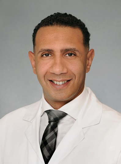
Dr. Mourad Abouelleil is a highly skilled and certified urologist renowned for his commitment to excellence in advanced urological procedures, including robotic prostatectomy and high-intensity focused ultrasound (HIFU). With state-of-the-art technology and extensive experience, Dr. Abouelleil offers personalized treatment plans tailored to meet the unique needs of each patient, ensuring high-quality medical care.
Error: Contact form not found.