
Nephrolithiasis, commonly known as kidney stone disease, affects approximately one in ten individuals during their lifetime. When mineral solutes in the urine—such as calcium oxalate, calcium phosphate, uric acid, or cystine—exceed their saturation threshold, they crystallize, aggregate, and form solid calculi within the renal collecting system. When these stones dislodge and migrate into the narrow ureter, they cause acute urinary obstruction, renal capsular distension, and the severe, colicky flank pain characteristic of renal colic.
Historically, the management of complex or obstructing kidney stones often necessitated open nephrolithotomy—a morbid procedure involving large, muscle-cutting flank incisions and surgical bisection of the renal parenchyma. Modern endourology has largely replaced open approaches with minimally invasive, endoscopic, and percutaneous modalities. Today, stone disease is treated along a tiered interventional spectrum, ranging from completely non-invasive acoustic shock waves to targeted percutaneous tract extraction.
In South Florida, Dr. Mourad Abouelleil is a board-certified urologist with the Palm Beach Health Network Physician Group, treating patients at Good Samaritan Medical Center in West Palm Beach and Palm Beach Gardens Medical Center. Having completed his medical training at the University of Wisconsin School of Medicine and Public Health and his urology residency at SUNY Upstate Medical University in Syracuse, New York, Dr. Abouelleil utilizes advanced endourologic technologies to resolve complex stone burdens. By matching intervention to stone volume, mineral density, and renal architecture, Dr. Abouelleil delivers complete stone-free clearance while prioritizing kidney preservation and rapid patient recovery.
Effective treatment planning requires detailed radiographic characterization of the stone and the surrounding urinary tract anatomy:
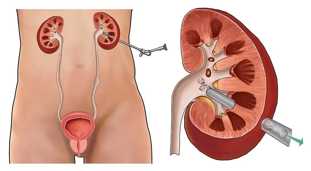
Dr. Abouelleil selects the appropriate intervention based on stone size, anatomical position within the kidney, and clinical urgency:
Extracorporeal Shock Wave Lithotripsy is the only completely non-invasive surgical modality for kidney stones:
Retrograde Intrarenal Surgery (RIRS) utilizes the body’s natural urinary openings without making any incisions:
For large, complex, or branched stones that exceed the safe working capacity of a ureteroscope, Percutaneous Nephrolithotomy provides definitive clearance:
| Clinical Metric | Shock Wave Lithotripsy (ESWL) | Ureteroscopy & Laser (URS/RIRS) | Percutaneous Nephrolithotomy (PCNL) |
| Surgical Invasiveness | Non-invasive (Zero incisions) | Endoscopic (Natural urinary orifice) | Minimally invasive (1 cm flank puncture) |
| Target Stone Size | Stones $< 1.5\text{ cm}$ | Stones up to $1.5\text{ to }2.0\text{ cm}$ | Large stones $> 2.0\text{ cm}$ / Staghorn |
| Stone Clearance Rate | 60% to 75% single-session | 85% to 92% single-session | 90% to 98% complete clearance |
| Anesthesia Type | Sedation or light general | General anesthesia | General anesthesia |
| Bleeding / Coagulation | Requires normal coagulation | Safe on active anticoagulation | Requires pausing anticoagulants |
| Lower Pole Clearance | Poor (gravity-dependent retention) | Moderate to high (with repositioning) | Superior direct-access clearance |
| Hospital Stay | Outpatient (Discharge in 1–2 hours) | Outpatient (Discharge same day) | Overnight observation (1 day) |
While most kidney stones are managed on an elective or urgent outpatient basis, one clinical scenario constitutes a critical urologic emergency: an obstructing stone accompanied by active urinary tract infection.
When bacteria proliferate behind an obstructing calculus, urinary stasis rapidly elevates renal pelvic pressures, forcing bacteria and endotoxins into the bloodstream via pyelovenous backflow. This can trigger life-threatening urosepsis and septic shock within hours.
Emergency Protocol: In the presence of fever, leukocytosis, and an obstructing stone on CT, definitive stone fragmentation is strictly contraindicated. The immediate priority is emergent urinary decompression. Dr. Abouelleil performs urgent retrograde ureteral stent placement or coordinates percutaneous nephrostomy tube placement to drain infected urine and stabilize the patient before scheduling definitive lithotripsy weeks later.
Treating existing kidney stones is only half of comprehensive management; preventing recurrence is critical, as up to 50% of stone formers develop another stone within five to ten years without dietary or medical prevention.
Following stone clearance, Dr. Abouelleil directs comprehensive metabolic evaluations:
Kidney stones do not follow a one-size-fits-all treatment path. From non-invasive shock wave therapy to advanced flexible laser ureteroscopy and percutaneous stone extraction, Dr. Mourad Abouelleil provides patients with targeted, minimally invasive options tailored to their anatomy, providing reliable stone-free outcomes while preserving kidney health.
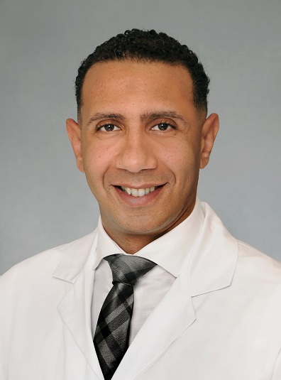
Dr. Mourad Abouelleil is a highly skilled and certified urologist renowned for his commitment to excellence in advanced urological procedures, including robotic prostatectomy and high-intensity focused ultrasound (HIFU). With state-of-the-art technology and extensive experience, Dr. Abouelleil offers personalized treatment plans tailored to meet the unique needs of each patient, ensuring high-quality medical care.
Error: Contact form not found.