The scrotum and testes serve two distinct yet intertwined biological functions: the endocrine production of systemic testosterone by Leydig cells and the exocrine production of spermatozoa within the seminiferous tubules. Pathologies affecting these compartments range from subtle systemic hormonal declines that impair metabolic vigor to acute or chronic fluid collections, varicoceles, and solid testicular tumors.
Because men often view scrotal enlargement, dull testicular discomfort, or progressive fatigue with anxiety or embarrassment, clinical evaluation is frequently delayed. Historically, scrotal complaints were evaluated primarily through physical examination alone, which carries high diagnostic error rates—particularly when differentiating between benign fluid-filled cysts and malignant parenchymal masses, or when evaluating the underlying etiology of secondary hypogonadism.
Modern urologic practice integrates targeted physical assessment with high-frequency Doppler ultrasonography and comprehensive endocrine laboratory profiling. In South Florida, Dr. Mourad Abouelleil is a board-certified urologist with the Palm Beach Health Network Physician Group, treating patients at Good Samaritan Medical Center in West Palm Beach and Palm Beach Gardens Medical Center. Having completed his medical degree at the University of Wisconsin School of Medicine and Public Health and comprehensive urology residency training at SUNY Upstate Medical University in Syracuse, New York, Dr. Abouelleil delivers structured diagnostic evaluations and minimally invasive surgical solutions to restore male endocrine balance and resolve scrotal disorders.
Evaluating Male Hypogonadism (Low Testosterone)
Male hypogonadism is defined as the failure of the testes to produce physiological levels of testosterone, normal sperm counts, or both. Clinical symptoms often manifest insidiously as chronic fatigue, diminished libido, erectile dysfunction, loss of lean muscle mass, depressed mood, and reduced bone mineral density.
1. The Diagnostic Laboratory Protocol
According to American Urological Association (AUA) guidelines, diagnosing testosterone deficiency requires strict biochemical confirmation:
- Fasting Morning Total Testosterone: Because serum testosterone exhibits circadian variation—peaking in the early morning hours—testing must be drawn between 7:00 AM and 10:00 AM after an overnight fast.
- Confirmatory Testing: A diagnosis cannot be made on a single blood draw; at least two separate morning tests documenting total testosterone levels $< 300\text{ ng/dL}$ are required.
- Free and Bioavailable Testosterone: In patients with conditions that alter sex hormone-binding globulin (SHBG)—such as obesity, diabetes, or liver disease—free testosterone provides an accurate measure of biologically active hormone.
2. Differentiating Primary vs. Secondary Hypogonadism
Determining the source of hormonal failure guides therapy:
- Primary Hypogonadism (Testicular Failure): Luteinizing Hormone (LH) and Follicle-Stimulating Hormone (FSH) are elevated, while testosterone is low. The pituitary gland is signaling appropriately, but the testicular Leydig cells cannot respond.
- Secondary Hypogonadism (Pituitary/Hypothalamic Failure): LH and FSH are low or inappropriately normal despite low testosterone levels. The defect lies in the brain’s signaling axis, which may warrant evaluating serum prolactin and obtaining pituitary MRI imaging if levels are markedly suppressed.
3. Therapeutic Pathways and Fertility Preservation
- Testosterone Replacement Therapy (TRT): Administered via intramuscular/subcutaneous injections, transdermal gels, or subcutaneous slow-release pellets. While TRT restores physiological androgen levels, exogenous testosterone suppresses the hypothalamic-pituitary-gonadal (HPG) axis, halting intratesticular spermatogenesis.
- Fertility-Preserving Alternatives: For men seeking to preserve active fertility or testicular volume, exogenous TRT is contraindicated. Instead, Dr. Abouelleil utilizes selective estrogen receptor modulators (SERMs, such as Clomiphene Citrate) or Human Chorionic Gonadotropin (hCG) to stimulate the body’s native LH receptors, prompting the testes to produce endogenous testosterone without suppressing sperm production.
Benign Scrotal Pathology: Hydroceles, Spermatoceles, and Varicoceles
Scrotal enlargement is most commonly driven by benign fluid accumulation or venous dilation rather than malignancy. High-resolution scrotal ultrasound with color Doppler is the gold-standard diagnostic tool:
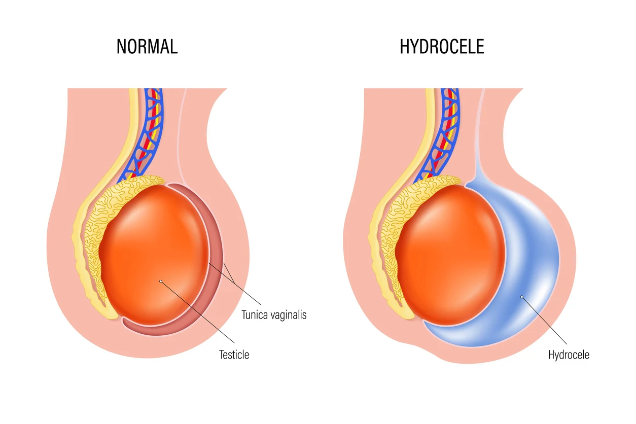
- Hydrocele: An abnormal accumulation of serous fluid between the parietal and visceral layers of the tunica vaginalis surrounding the testis. It presents as a painless, fluctuant scrotal enlargement that transilluminates under a focused light source. While small hydroceles can be observed, large, tense hydroceles that cause chronic dragging discomfort or cosmetic distress are treated with an outpatient hydrocelectomy (excision and eversion of the tunica vaginalis sac).
- Spermatocele (Epididymal Cyst): A benign, fluid-filled cystic structure originating from the efferent ductules of the epididymal head, typically containing non-viable sperm. They feel like a discrete, smooth, mobile marble distinct from the testicle itself. Surgical excision is indicated only if the spermatocele becomes painful or reaches symptomatic size.
- Varicocele: An abnormal tortuosity and dilation of the pampiniform venous plexus within the spermatic cord, colloquially described as feeling like a “bag of worms.” Because the left testicular vein empties into the left renal vein at a perpendicular angle, over 85% to 90% of varicoceles occur on the left side. Varicoceles elevate scrotal temperature, which can impair spermatogenesis and Leydig cell function. When associated with male-factor infertility or persistent dull aching, microsurgical subinguinal varicocelectomy provides definitive venous ligation.
Comparative Overview: Diagnostic Presentation of Common Scrotal Masses
| Condition |
Anatomical Location |
Physical Exam Characteristic |
Scrotal Ultrasound Appearance |
Primary Clinical Management |
| Hydrocele |
Surrounds entire testicle |
Fluctuant, soft; positive transillumination |
Anechoic (clear fluid) surrounding testis |
Observation or Hydrocelectomy |
| Spermatocele |
Distinct; head of epididymis |
Smooth, mobile nodule superior to testis |
Well-defined cystic lesion separate from testis |
Observation or Spermatocelectomy |
| Varicocele |
Spermatic cord (Upper scrotum) |
“Bag of worms”; distends during Valsalva |
Dilated veins $> 3\text{ mm}$ with retrograde Doppler flow |
Microsurgical Subinguinal Ligation |
| Testicular Cancer |
Within testicular parenchyma |
Hard, painless, non-transilluminating rock |
Hypoechoic, vascularized intratesticular mass |
Radical Inguinal Orchiectomy |
The Workup of a Solid Testicular Mass: Rule Out Malignancy
Any firm, hard, or painless intratesticular mass must be treated as a potential testicular germ cell tumor until proven otherwise. Testicular cancer is the most common solid malignancy in young men aged 15 to 35, though it carries cure rates exceeding 95% when identified promptly.
1. High-Frequency Scrotal Ultrasonography
The initial diagnostic branch point is determining whether a palpable mass is extratesticular (arising from the epididymis or spermatic cord, which is almost always benign) or intratesticular (arising within the testicle itself, where 90% to 95% represent malignancy). A solid, hypoechoic, hypervascularized mass within the testicular parenchyma demands immediate oncologic evaluation.
2. Serum Tumor Markers
Prior to any surgical intervention, baseline serum tumor markers must be drawn to assist in staging and histological classification:
- Alpha-Fetoprotein (AFP): Elevated in non-seminomatous germ cell tumors (specifically embryonal carcinoma and yolk sac tumors); never elevated in pure seminoma.
- Beta-Human Chorionic Gonadotropin (Beta-hCG): Elevated in choriocarcinomas and a subset of pure seminomas.
- Lactate Dehydrogenase (LDH): A non-specific marker reflecting overall tumor burden and cellular turnover.
3. The Surgical Cardinal Rule: Radical Inguinal Orchiectomy
If an intratesticular mass is confirmed on ultrasound:
- Percutaneous Biopsy is Strictly Contraindicated: Performing a needle biopsy through the scrotum is prohibited because it breaches local anatomical lymphatic barriers. The scrotum drains into superficial inguinal lymph nodes, whereas the testicle drains retroperitoneally into lumbar lymph nodes. A trans-scrotal needle puncture can disrupt these planes, altering the metastatic pathway.
- Radical Inguinal Orchiectomy: Dr. Abouelleil performs surgical extirpation via a small transverse groin incision in the inguinal canal. The spermatic cord is clamped at the internal inguinal ring before the testicle is mobilized, delivering the intact testicle and high cord for definitive histopathological staging. A cosmetic silicone testicular prosthesis can be placed simultaneously if desired.
Conclusion & Practice Information
Whether managing systemic hormonal deficiencies, uncomfortable scrotal fluid collections, or a concerning testicular nodule, structured diagnostic evaluations ensure clear answers and targeted care. Through clinical hormonal management, in-office ultrasound assessments, and minimally invasive microsurgical and reconstructive techniques, Dr. Mourad Abouelleil helps men resolve scrotal disorders and preserve long-term endocrine vitality.
- Surgeon: Dr. Mourad Abouelleil, MD
- Board Certification: American Board of Urology
- Practice Group: Urology Medical Associates – Palm Beach Health Network Physician Group
- Hospital Affiliations: Good Samaritan Medical Center (West Palm Beach) & Palm Beach Gardens Medical Center
- Primary Office Locations:
- West Palm Beach: 1411 North Flagler Drive, Suite 3800, West Palm Beach, FL 33401
- Jupiter: 641 University Boulevard, Suite 105, Jupiter, FL 33458
- Port St. Lucie: 900 Southeast Becker Road, Port St. Lucie, FL 34984
- Office Consultations: (561) 291-7182