For men diagnosed with localized prostate cancer, surgical removal of the prostate gland remains one of the most reliable pathways toward long-term cancer eradication. However, the male pelvis is a narrow, rigid anatomical corridor where oncologic control and functional quality of life are closely linked. The microscopic neurovascular bundles that govern erectile function travel directly along the outer surface of the prostate capsule, while the urinary sphincter wraps tightly around the prostatic apex.
Historically, traditional open radical prostatectomy required large lower-abdominal incisions and was frequently complicated by significant blood loss, prolonged hospital stays, and variable recovery of urinary continence and erectile potency.
The advent of Robotic-Assisted Radical Prostatectomy (RARP) transformed this operation into a precise, minimally invasive procedure. Utilizing high-magnification 3D stereoscopic vision, tremor-filtration software, and wristed micro-instruments, surgeons can navigate complex pelvic anatomy with sub-millimetre precision.
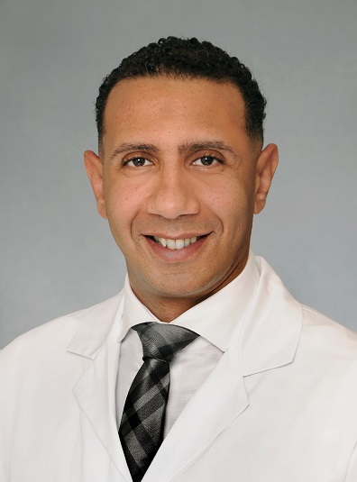
In South Florida, Dr. Mourad Abouelleil is a board-certified urologist with the Palm Beach Health Network Physician Group, serving patients at Good Samaritan Medical Center in West Palm Beach and Palm Beach Gardens Medical Center. Having completed his medical degree at the University of Wisconsin School of Medicine and Public Health, followed by extensive residency training in general surgery and urology at SUNY Upstate Medical University, Dr. Abouelleil has performed thousands of robotic and minimally invasive urologic surgeries. He employs advanced, patient-tailored nerve-sparing protocols to completely resect localized prostate cancer while preserving the delicate neurovascular and sphincteric anatomy responsible for urinary control and sexual vitality.
The Functional Anatomy: Neurovascular Bundles and Sphincteric Complex
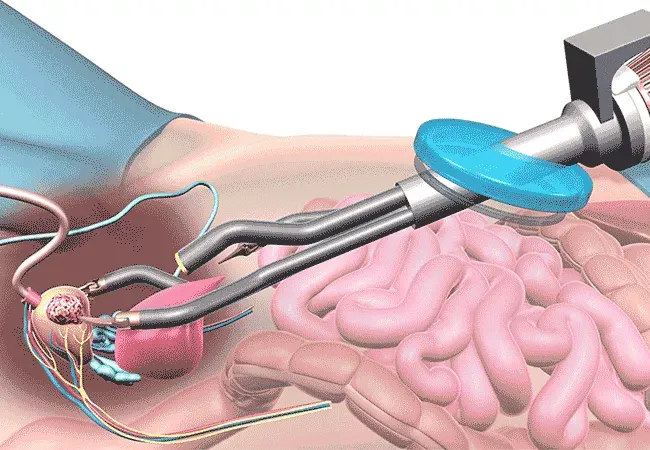
Preserving post-prostatectomy function requires an in-depth understanding of the pelvic fascial planes:
- The Cavernous Nerve Plexus: Erectile function is mediated by autonomic parasympathetic and sympathetic fibers that emerge from the pelvic plexus. These microscopic nerves travel within a dynamic meshwork—the neurovascular bundle (NVB)—encased between the prostatic fascia and the levator ani fascia along the posterolateral surface of the prostate.
- The External Striated Sphincter (Rhabdosphincter): Located at the prostatic apex, this omega-shaped collar of striated muscle fibres wraps around the membranous urethra. Because the internal urinary sphincter located at the bladder neck is removed during surgery, preserving the length, innervation, and vascular supply of the external sphincter is paramount for achieving early urinary continence.
Graded Nerve-Sparing Protocols: Matching Biology to Dissection Planes
Nerve sparing is not an “all-or-nothing” decision. Dr. Abouelleil utilizes a graded fascial approach, selecting the dissection plane based on the patient’s baseline erectile function, multiparametric MRI findings, and the location of positive biopsy cores:
- Intrafascial Dissection (Full Nerve Sparing): The surgical dissection plane is established directly along the true prostatic capsule, beneath the prostatic fascia. This preserves virtually all neural and vascular tissue within the bundle. It is reserved for low- to intermediate-risk tumours with no evidence of capsular abutment or extraprostatic extension (EPE).
- Interfascial Dissection (Partial Nerve Sparing): Dissection proceeds between the layers of the prostatic fascia and the levator fascia, taking a thin protective cushion of tissue while leaving the majority of the outer neurovascular bundle intact. This approach is selected when the tumor approaches the capsule, balancing margin safety with functional preservation.
- Extrafascial Dissection (Wide Resection / Non-Nerve Sparing): The plane is taken outside the levator fascia, completely resecting the neurovascular bundle along with the prostate. This is reserved for aggressive, high-risk disease where cancer has demonstrably breached the prostate capsule, prioritizing complete oncologic clearance.
- Incremental / Asymmetric Sparing: Because prostate cancer is often asymmetric, Dr. Abouelleil can perform full intrafascial nerve sparing on the unaffected side while performing a wider, protective resection on the side harbouring the index tumour.
Surgical Execution: The Steps of a Nerve-Sparing Robotic Prostatectomy
The procedure is performed through five to six small keyhole incisions (5 to 12 mm) across the abdomen, utilizing the da Vinci surgical system:
- Developing the Retropubic Space: The peritoneal cavity is accessed, and the bladder is gently mobilized anteriorly to expose the space of Retzius, preserving the anterior pelvic support structures.
- Seminal Vesicle and Vas Deferens Mobilization: The vas deferens are divided, and the seminal vesicles are dissected athermally, releasing Denonvilliers’ fascia from the anterior rectal wall.
- Athermal Pedicle and Nerve-Bundle Release: Rather than using monopolar or bipolar electrocautery near the neurovascular bundles—which can cause thermal neuropraxia (nerve stun)—delicate vascular pedicles feeding the prostate are controlled using titanium micro-clips or precise hemoligation, peeling the nerve bundles away without heat injury.
- Apical Dissection and Urethral Preservation: Under high 3D magnification, the dorsal venous complex is controlled, and the junction between the prostatic apex and membranous urethra is sharply divided, preserving maximal functional urethral length to promote urinary continence.
- Pelvic Lymph Node Dissection: For intermediate- and high-risk disease, an extended or standard pelvic lymphadenectomy is performed, clearing the obturator and external iliac nodal packets for accurate staging.
- Watertight Vesicourethral Anastomosis: A running, continuous micro-suture connects the bladder neck to the preserved urethral stump, creating a secure, watertight seal over a silicone Foley catheter.
Comparative Overview: Open Surgery vs. Robotic-Assisted Prostatectomy
| Clinical Metric |
Traditional Open Radical Prostatectomy |
Robotic-Assisted Radical Prostatectomy (RARP) |
| Incision Size |
8 to 12 cm midline lower abdominal incision |
5 to 6 keyhole ports (5–12 mm each) |
| Surgical Visualization |
Standard visual field; deep, shadowed pelvis |
10x to 15x 3D high-definition magnification |
| Blood Loss (EBL) |
500 to 1,500+ mL (higher transfusion rates) |
Minimal (< 50 to 100 mL; transfusions rare) |
| Tissue Handling |
Rigid instruments with limited pelvic reach |
EndoWrist® instruments with 7 degrees of freedom |
| Catheter Duration |
Typically 10 to 14 days |
Typically 6 to 8 days post-surgery |
| Hospital Stay |
2 to 4 days inpatient |
Overnight observation (1 day discharge) |
| Return to Normal Activity |
6 to 8 weeks |
2 to 3 weeks |
Functional Recovery: Continence and Potency Pathways
Achieving the “trifecta” of prostate cancer surgery—negative margins, continence, and potency—depends on both surgical technique and postoperative rehabilitation:
- Urinary Continence: The majority of patients experience transient stress leakage after catheter removal as the external sphincter adapts. Continence recovery is supported by pre- and postoperative pelvic floor muscle exercises (Kegels). With maximal urethral length preservation, most men achieve zero-pad or single-security-pad control within weeks to a few months.
- Erectile Rehabilitation: Because peripheral nerves heal slowly (approximately 1 mm per day), cavernous nerves require time to recover from surgical manipulation. Dr. Abouelleil initiates an active penile rehabilitation program shortly after catheter removal, utilizing daily low-dose PDE-5 inhibitors (e.g., Tadalafil) to promote continuous arterial oxygenation and prevent corporal fibrosis during the neurological recovery period.
Conclusion & Practice Information
Robotic-assisted radical prostatectomy provides a refined balance between definitive cancer cure and functional preservation. Through tailored nerve-sparing dissection, athermal tissue handling, and maximal urethral length preservation, Dr. Mourad Abouelleil offers patients an evidence-based pathway designed to eliminate localized prostate cancer while protecting the urinary and sexual functions that matter most.
- Surgeon: Dr. Mourad Abouelleil, MD
- Board Certification: American Board of Urology
- Practice Group: Urology Medical Associates – Palm Beach Health Network Physician Group
- Hospital Affiliations: Good Samaritan Medical Center (West Palm Beach) & Palm Beach Gardens Medical Center
- Primary Office Locations:
- West Palm Beach: 1411 North Flagler Drive, Suite 3800, West Palm Beach, FL 33401
- Jupiter: 641 University Boulevard, Suite 105, Jupiter, FL 33458
- Port St. Lucie: 900 Southeast Becker Road, Port St. Lucie, FL 34984
- Office Consultations: (561) 291-7182